
Lorenzo Tei
UNIUPO, PA Chim / 06: Supervisor of the preparation and characterization of nanosystems. Responsible for organic synthesis.
Read bio
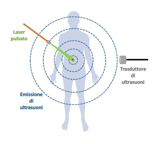
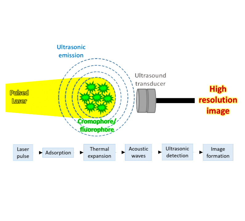
The innovative photoacoustic imaging (also known as optoacoustic imaging) exploits the ability of chromophores/fluorophores, duly excited by light, to emit mechanical energy in the form of ultrasounds. The ultrasounds, detected by appropriate piezoelectric, are used to generate high-resolution images capable of providing biomedical, structural and functional information on specific anatomical regions of interest.

Patent status
GRANTED

Priority Number
102016000023103

Priority Date
04/03/2016

License
ITALIA
Market
TAM - Diagnostic imaging analysis of tumors, through the use of contrast agents SAM - Market for photoacoustic imaging contrast agents in oncological diseases, such as breast cancer and melanoma. SOM - market for photoacoustic imaging contrast agents for preclinical use in animal models
Problem
In the field of diagnostic imaging, a new diagnostic method called Photoacoustic Imaging (PAI) has developed over the last decade. This technique, currently very widespread at the preclinical level, is starting to find its first applications in the clinical field (eg breast cancer study, finger vascularity analysis, melanoma study, ...). The contrast can be generated by endogenous molecules (eg hemoglobin, melanin, ...) or by exogenous molecules (eg fluorochromes, metal nanosystems, ...). The development of new and efficient exogenous contrast agents is therefore central to the development of the method and its diffusion in the preclinical and clinical setting. With the goal of a rapid development of contrast agents, organic fluorescent molecules, already approved for use in patients, such as indocyanine green (ICG), were explored. In fact, this molecule is widely used in the clinic, for example for angiography of the retinal vessels or for the analysis of sentinel lymph nodes. Two common problems related to the use of ICG are: i) the strong degradation in the physiological environment, by oxygen or other factors of the microenvironment, ii) the easy in vivo sequestration by albumin with consequent rapid elimination, iii) the non-specific biodistribution. There is therefore the need to identify new contrast agents for photoacoustic applications. Up to now, there is NO contrast agent used in the medical clinic in the photoacoustic field. Hence this is a field of considerable economic importance destined to expand.


Current technology limitations
Diagnostic imaging is increasingly assuming a central role in medical practice, allowing a more detailed and fast diagnosis. This results in an improvement of therapeutic performance. Even in the preclinical field, diagnostic imaging is playing a key role, allowing, in a non-invasive way, to identify physiological and pathological events and to rapidly test new therapeutic approaches. Various imaging techniques are currently available, such as MRI, PET / SPECT, CT, US, OI. Photoacoustic imaging is a relatively new technique, with some intrinsic advantages over other imaging methods, namely: i) the absence of invasiveness and toxicity for the patient and ii) the ease and speed of execution (not requiring complex technologies or expensive). Currently, the photoacoustic imaging method is in the early stages of clinical application and suitable contrast agents have NOT yet been developed. Some potential contrast agents are already on the market such as, for instance, Indocyanine Green (ICG). Unfortunately, they have some limitations of applicability related mainly to i) rapid elimination from the blood (low plasma half-life), ii) rapid oxidative degradation and iii) use only as a passive agent. In this sense, in the oncology field, the ICG passively accumulates in the tumor due to the EPR effect (enhanced permeability and retention). The encapsulation of the ICG in biocompatible systems such as mesoporous silica (MSNs) allows to overcome all these limits, expanding the potential of the dye in the photoacoustic field. In addition, the contrast generated by the ICG encapsulated within the MSNs is greater than that one generated by an equal dose of ICG administered in free form. Therefore, another advantage of this system is the possibility to reduce the dose of ICG to be administered to the patient.
Killer Application
The first possible application would be in melanoma diagnostics. This tumor represents one of the most aggressive human cancers with a high mortality rate (melanomapatients.org.au). Early diagnosis and follow-up of therapies significantly increase the chances of therapeutic success. A negative prognostic factor is represented by the high degree of vascularization and high vascular permeability. The use of photoacoustic imaging, with the addition of our product, can be of great help in the early diagnosis of this tumor and in its phenotyping, allowing to evaluate the vascular parameters of interest for tumor monitoring. In fact, our contrast agent would allow us to evaluate i) tumor margins, ii) the degree of vascularization and iii) vascular perfusion. All this would be added to the information obtainable by photoacoustic imaging and ultrasound without contrast, namely i) tumor morphology, ii) vascular volume and hypoxia, iii) content of melanin.

Our technology and solutions
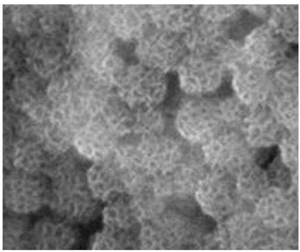
The product is a novel photoacoustic imaging contrast agent (PAI). This product is made by mesoporous silica nanosystems containing the ICG fluorescent dye inside. The preparation can be freeze-dried and stored in dried form, to be resuspended in physiological saline immediately before use. The encapsulation of the ICG in biocompatible systems, such as mesoporous silica (MSNs), allows to overcome the limitations normally associated with the use of ICG in free form, expanding the potential of the dye in the photoacoustic field. In addition, the contrast generated by the ICG encapsulated within the MSNs is greater than that one generated by an equal dose of ICG administered in free form. Therefore, another advantage of our system is to allow the reduction of dose of ICG to be administered to the patient. Administration to the patient can take place via intravascular injection, immediately before carrying out the photoacoustic examination. Photoacoustic scans are performed before administration of the contrast agent (pre images) and after administration, at variable times. In this way it is possible to follow the kinetics of accumulation of the probe in the tumor site, allowing to extrapolate physiological information on the tumor such as the vascular volume and the permeability of the vessels. This provides information on the nature of the tumor. The main field of use can be that of melanomas and breast tumors, since they are superficial tumors easily reachable by the photoacoustic imaging technique (PAI).
Advantages
The developed product would represent the first contrast agent for photoacoustic imaging with possible use in oncology. In particular, the photoacoustic imaging technique, in which our product is applied, has some intrinsic advantages in comparison to other imaging methodologies such as magnetic resonance imaging (MRI) or nuclear medicine (PET / SPECT), i.e. : i) the absence of invasiveness and toxicity for the patient and ii) the easy and fast execution (not requiring complex or expensive technologies). The use of contrast agents, such as the one we propose, allows to add these advantages of the imaging method to the possibility of obtaining physiological and metabolic information of the tumor (vascular volume, permeability of the vessels, perfusion, etc ...). Two more elaborate versions of the product could i) contain 'targeting' molecules on the surface, which mediate specific accumulation at the tumor site and ii) be filled not only with fluorescent dye but also with a drug, to combine diagnostics and therapy (teranostic system).
Roadmap
The study will continue with the analysis of the biodistribution of the nanosystem and the analysis of biocompatibility, performed on a sufficient number of animals. In this way, LD50, ED50, clearance, plasma half-life time will be evaluated Following, the idea is to contact the 2i3T University Business Incubator to check the feasibility of technology transfer, to efficiently assess the development potential of the idea and to assess which are the potential market players and the key elements for transferability. This will serve to define the most suitable know-how enhancement mechanism, allowing us to evaluate between the possibility of IP transfer (most likely hypothesis) or the possible establishment of an innovative Start-up
TRL
Il team