

Clizia Chinello
Clizia Chinello, PhD expert in proteomics. Leader of proteome identification and characterisation by nano LC-MS / MS group.
Read bio
Since 2012 the UNIMIB Clinical Proteomics and Metabolomics Unit in collaboration with the Anatomia Patologica group has led not only to numerous publications in the most important sector journals and to the success of some competitive scholarships (FP7-PEOPLE-2013-ITN, AIRC MFAG 2016, PRIN 2017) also to the filing of a patent concerning a diagnostic method for the recognition of the “molecular signature” of malignant thyroid neoplasms (consisting of a combination of proteins expressed by tumor cells and not by benign nodules) based on proteomics through advanced mass spectrometry (MALDI-TOF; MS-Imaging). It allows a diagnosis of cancer on the same biopsy sample in which the conventional pathological analysis is indeterminate. Currently, after consolidating the methodological aspects, removing the interference due to the high amount of hemoglobin and stabilizing the biological material for at least 10-14 days in order to allow centralized analysis with sampling from different clinical centers, more than 200 samples. The statistical algorithm that discriminates between malignant and benign is being optimized with over 100 patients for the educational phase and another 100 patients for the verification phase. Within a few months we will have the definitive model for the subsequent clinical validation phase.

Patent Status
GRANTED

Priority Number
102015000062301

Priority Date
16/10/2015

License
ITALY

Market
EPIDEMIOLOGY
The incidence of palpable nodules is 3-5% of the general
population, whilst US-ecographic lesions are detected in
approximately 20-75% of the general population.
The cancer incidence per year is 0.1%;
around 0.5.-10 cases per 100,000
Thus, around 5% of nodules are cancer.
These epidemiologic data strongly a big addressable market.
Problem
The pre-operative distinction between benign and malignant thyroid conditions is essential to avoid unnecessary treatment of patients and morbidity due to inappropriate surgery. The most widely used tool for making a diagnosis is the ultrasound guided needle aspiration (FNA). Unfortunately, a significant number of FNAs (20-30%) are "indeterminate to malignancy" (TIR3) after the traditional pathological examination under an optical microscope, representing an important problem for these patients whose thyroid is surgically removed for a diagnostic, and not curative, purpose.
There are currently no alternatives to fine needle aspiration (FNA) biopsies which are universally recognized as the procedure of choice in the preoperative evaluation of thyroid nodules.


Current Technology Limits
The pre-operative distinction between benign and malignant thyroid conditions is essential to avoid unnecessary treatment of patients and morbidity due to inappropriate surgery. The most widely used tool for making a diagnosis is the ultrasound guided needle aspiration (FNA). Unfortunately, a significant number of FNAs (20-30%) are "indeterminate to malignancy" (TIR3) after the traditional pathological examination under an optical microscope, representing an important problem for these patientswhose thyroid is surgically removed for a diagnostic, and not curative, purpose.
There are currently no alternatives to fine needle aspiration (FNA) biopsies which are universally recognized as the procedure of choice in the preoperative evaluation of thyroid nodules.
Killer Application
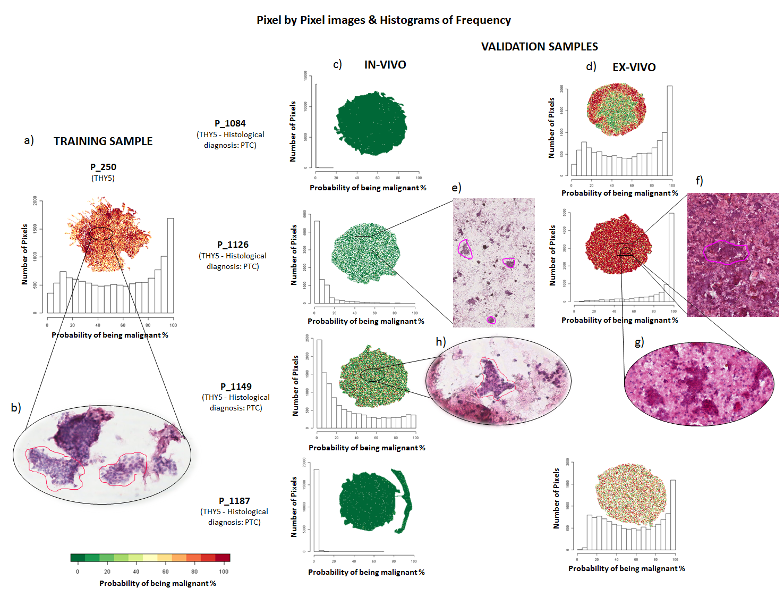
The starting product, consisting of molecular signatures capable of differentiating benign from malignant lesions, can constitute the starting point for building subsets of specific signatures for the different histotypes of the lesions. Furthermore, and perhaps even more importantly, it can also be used to develop those capable of detecting the presence of pre-cancerous forms, even more difficult to diagnose by the pathologist, that, when not detected, constitute a potential high-risk population.

Our Technology and Solutions
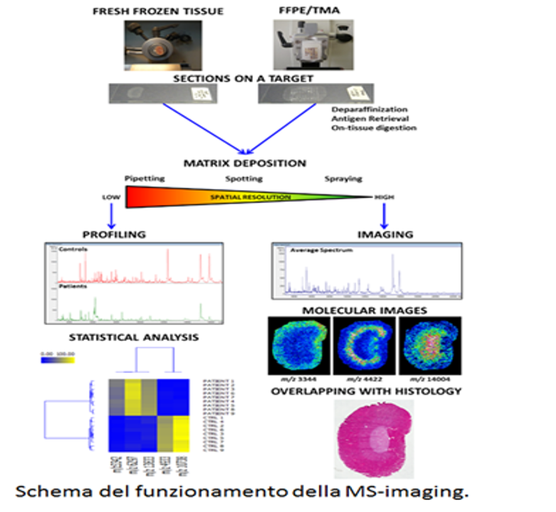
A possible alternative is offered by a proteomic approach in which an imaging technique using mass spectrometry (MALDI-imaging mass spectrometry) is used to identify molecular signatures at the protein level.
Mass spectrometry imaging (MSI) generates thousands of images for each type of analyte, thus offering a molecular dimension to routine histopathology. MSI is able to produce molecular images of biopsy findings, complementary to those of microscopy or immunofluorescence, but more informative as it highlights the alterations even of the single cell and therefore provides specific molecular signatures of normal or "altered" cells. This attribute (resolving the spatial localization of multiple proteins within a single section of pathological tissue) enables the pathogenetic molecular
"actors" to be identified, improving the understanding of thyroid pathology and can be translated into clinical tools for patient care.
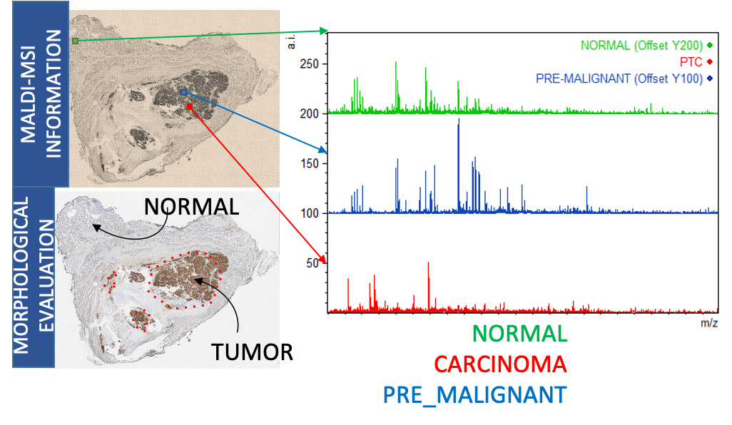
The purpose of the present invention is therefore to provide a method for the in vitro differential diagnosis of thyroid pathologies based on the use of specific molecular signatures of malignant and benign thyroid lesions, obtained by
MALDI-MSI, capable of correctly classifying the type of lesion.
Advantages
- Reduce the number of unnecessary thyroidectomies
- Improve the diagnosis of thyroid lesions
- Reduce the discomfort for patients due to unnecessary removal of the thyroid
- Reduce the economic strain placed on the NHS due to surgical interventions and the consequent life-long drug treatment

Roadmap
The product can be marketed in various ways:
- selling the use of molecular signatures to MS companies for use in the clinic
- sale of molecular signatures to MS companies for use in the clinic
- sale of molecular signatures to software-houses for the generation of diagnostic applications to be associated with MS
TRL
The Team