
Viviana Frantellizzi
Researcher in Diagnostic Imaging at the Sapienza University of Rome
Nuclear Doctor at the Policlinico Umberto I in Rome. He has published numerous articles in international impact magazines.
Read bio
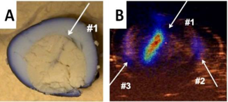
The invention relates to an echoscintigraphic probe for medical applications and the related procedure for images fusion. It is based on an ultrasound probe integrated with a scintigraphic device or gamma camera. This prototype provides fused images revealing both anatomical and functional details. The ultrasound probe is housed on the top, above the collimator plane and hangs out to facilitate the contact with the patient body. The collimator is able to obtain images of the radiolabeled drug biodistribution, preserving the ultrasound probe characteristics. The probe is applicable to intra-operative clinical applications and to diagnosis of tumor pathologies by means of radioactive tracers.


Patent Status
GRANTED

Priority Number
102014902261930; EP3143428; US10638997

Priority Date
16/05/2014

License
INTERNATIONAL

Market
The ecoScinti is a highly innovative tool and unique, therefore making it difficult to accurately analyze and quantify market potential. “Assobiomedica” census (Nov. 2015) reports that there are just over 32,000 ultrasound scanners in Italy. The period of “technological suitability” of an ultrasound machine is 5 years, and only 50% of the instruments in Italy fall within this range. Just over 10% of the instruments currently in operation are even over 10 years old and the life expectancy is 6 years. Given these rather long life cycles, we believe that the development of an echo-scintigraphic probe that can also be used by instruments already in use is the strategy that will allow us to achieve the widest and fastest penetration of the market. Considering the highly innovative characteristics of the device, together with the sales costs lower than those of the main equipment currently used for dual-imaging, it is plausible to assume a good diffusion of the instrument. We estimated the potential market considering the number of existing nuclear medicine (NM) centres, starting point for the inoculation of the radiotracer of any scintigraphic analysis: ITALY ~250 NM, EUROPE >2,500 NM, USA >5,000 NM. The following eco-numbers of scintigraphic probes sold in the first three/four years from the completion of the clinical study and the placing on the respective markets can be hypothesized: ITALY 600 units, EUROPE 6,000 units, USA 12,500 units. Placing the price of our probe at around €40,000 per unit, the turnover numbers in four years would be: ITALY €24 million, EUROPE €240 million, United States €500 million. With these sales numbers, gross margin could be 65-70% of revenues.
Problem
To date modern diagnostic, interventional and clinical medicine need a technological improvement in the field of imaging that combines anatomical and functional information in the study of an organ. Since the scintigraphic images do not provide anatomical information, but only functional, to solve the problem SPECT and PET are normally combined with CT. Several attempts have been made to merge the SPECT/PET images with the anatomical information of ultrasound, but all carried out using complex methods that need separated imaging systems: a software system that combines the two images and an external optical tracking system (mainly invasive) to correlate spatial information coming from the two instruments. However, the complexity of use, the persistence of the dissociation of the methodical and the inaccurate overlapping have meant that the fusion attempts of SPECT/PET images with the anatomical details of the ultrasounds have not yet been applied in clinical practice. To answer the need for an ultrasound instrument that integrates metabolic information, We intend to bring to market the first eco-scintigraphic probe: a probe for ultrasound and scintigraphy imaging with merging of the images in real time, without the need for bulky and expensive additional tools for correlation. The images, superimposed in real-time, one-shot, provide different information: • anatomical details detected by ultrasound to help to identify lesions in high-definition • functional image, obtained from a gamma detector array, used to characterize the lesion and its metabolism with extreme precision, thanks to the millimetric spatial resolution. The combination of the two images will improve the diagnosis and metabolic characterization of many diseases.
Current Technology Limits
Recently, several studies have demonstrated the great capacity of “dual-imaging“ diagnostic systems, systems that are able to provide a double image of the object under examination. The basic criterion is to combine two complementary images: morphological image that provides anatomical details (RX, CT and NMR) with another image that can provide information about the biological function (images obtained using radio tracers, such as PET and SPECT ). The proposed solutions are huge systems based on the use of a tomographic ring in which two detectors are integrated as SPECT CT, PET CT, CT NRM and most recently PET NRM. All these systems have the disadvantage of being extremely bulky and very expensive, and therefore are an exclusive prerogative of large industrial groups such as General Electric, Siemens, Philips and Toshiba, which produce similar systems with small differences from each other. There have been many research projects to integrate ultrasound images with SPECT images, but none has proved to be industrially and commercially viable. None solution was found to be a real solution, for the need of external correlation systems to superimpose images or for lack of a real-time overlapping. To date the current market of a dual-imaging handheld instrument is therefore devoid of competitors, while the solutions offered by large industrial groups are products very different.
Killer Application
Our Technology and Solution
The dual modality probe is based on an UltraSound (US) detector integrated with a small Field of View (FoV) single photon emission gamma camera. The great innovation of this probe is in its handheld configuration and in the perfect coregistration of dual images. The gamma detector is based on a slit collimator that, thanks to its high efficiency, allows to obtain images in few seconds (the acquisition time is close to the one of an US examination). The perfect alignment between US and gamma devices is established through the alignment of the US piezoelectric strip with the inter-spaces of the collimator slit guaranteeing the perfect overlap between US and gamma images.
Advantages
The strong impact all over the world can be evaluated taking into account the number of patients subjected to diagnostic treatments (about 10 million people for year in Europe and 20 million in USA). Since this device can be used on all patients needing to perform diagnostic tests, the catchment area is about 30 million of people.
Roadmap
Considering the excellence of innovation of the instrument and its high social-economic impact, we are confident that the project has the features to get appropriate contributions from the European institutions (H2020) and/or national or regional funding.
TRL
Team