This medical device for melanoma screening can characterize and evaluate the potential risk of nevi based on data gathered through Structure-from-Motion (SfM) digital photogrammetry and infrared thermography. Thanks to Artificial intelligence the device associates a risk factor index to each detected skin lesion, thanks to the deep learning based on quantitative and qualitative information, three dimensional morphology, chromatic and thermographic data as well as the variation of such measurements over time.

Patent Status

PENDING

Priority Number

102016000132357

Priority Date

29/12/2016

License

INTERNATIONAL

Market

Melanoma is the most aggressive skin cancer. Its incidence has increased dramatically over the past three decades. The importance of early screening is paramount: according to Industrydataanalytics.com, the dermatoscope market alone could witness a revenue growth of 10.2% CAGR, for a global market value of USD 53M by 2026. This medical device targets public and private hospitals and clinics specialized in dermatology, estimating an early adopter market of about 400-500 pcs. The technology will therefore be ideally marketed by manufacturers of medical imaging systems (medical devices, dermatoscopes, diagnostic devices, skin cancer, melanoma screening, etc.).

  • TAM: USD 19.43 bn in 2023
  • SAM: estimated at USD 9.09 bn in 2023
  • SOM: estimable between USD 9.09 bn and USD 18.18 bn in 2023

Problem

Currently, the diagnosis of skin cancer is characterized by a high degree of subjectivity, due to the impossibility to perform an accurate analysis of all nevi in the limited time of a single visit. This increases the risk of a failure to timely detect any suspicious melanoma. The human eye is in fact limited and does not allow a precise and quantitative analysis of the typical features of melanocytic nevi.  Consequently, due to an excess of caution, many nevi are surgically removed unnecessarily: in fact, it is estimated that only 20% of biopsies performed  result in a positive melanoma diagnosis. Current technologies, based on visual analysis and dermoscopy, cannot accurately monitor the evolution of the patient’s skin over time, which is a key aspect of the medical evaluation.

Moreover, the technologies currently available to dermatologists for the screening of melanomas and other skin diseases are not able to:

  • Analyze a large number of nevi (at times hundreds) in a short time
  • Make an evaluation based on objective parameters
  • Make an accurate comparison of changes over time
  • Combine information collected by different diagnostic tools

Current Technology Limits

At present, no existing equipment can implement all the solutions and outputs that this patented system provides in a unified manner. There are systems that provide three-dimensional skin data, but do not include thermographic data and do not provide a classification of nevi according to a risk index. There are also systems that provide classification of nevi, but are limited to two-dimensional images in unpolarised light. Furthermore, the output formats of such examinations often require proprietary software and are therefore not interoperable.

Today, the diagnosis of cutaneous melanomas is therefore characterised by a high degree of subjectivity, precisely because these instruments are unable to make accurate measurements of many nevi in the limited time of an examination. Since they are based exclusively on visual analysis and dermoscopy, screening is not optimal because it does not take into account the evolution of the patient’s skin over time.

The evaluation of whether or not to perform a biopsy therefore depends largely on the experience of the dermatologist, who does not have a support of objective and automated data output. Biopsies – or other diagnostic methods – may therefore be negative (in the case of analysis of melanomas that are not actually suspicious), partial or late (in the case of suspicious nevi that are not detected in due time).

Killer Application

This medical instrument will become an indispensable tool to support dermatologists in detecting suspicious nevi affected with melanoma, as well as in the periodic patient screening. The technology is therefore mainly addressed to manufacturers of medical diagnostic imaging systems and equipment, with reference to both the hardware system (digital cameras, thermal imaging cameras) and the software architecture accompanying the technology. Further developments of the technology could involve the diagnosis of other skin diseases.

Our Technology and Solution

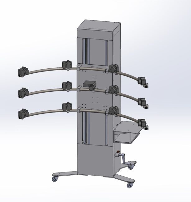
The prototype currently in use for experimental clinical validation at the Istituto Oncologico Veneto Cancer Institute, is able to acquire data related to the skin of the patient’s back, thanks to the data provided by 12 synchronised cameras (operating in polarised light) and a thermal camera, positioned on a curved and mobile frame.

The calculation of the risk factor index associated with each nevus is achieved through the work of convolutional neural networks (CNN) that classify the information acquired during screening on the basis of the information acquired during training and comparison with previous screenings.

The data acquired include:

  • Photographic data;
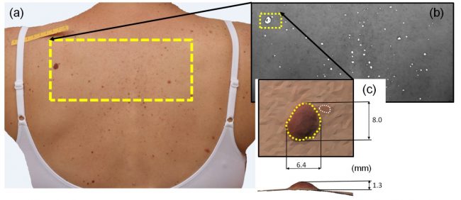
  • Photogrammetric modelling, which returns the nevi geometry obtained from polarised light images with 0.2mm resolution;
  • Fusion of chromatic and thermal data;
  • Correlation of the thermographic data with the level of vascularisation;
  • Comparison with screening from previous examinations to detect variations.

The system provides an output in DICOM format:

  • The 2D model of the skin with indication of the melanoma risk factor index for each skin lesion detected
  • The set of data that characterise the nevi identified as having the highest risk factor.

The DICOM format is universally used for the management of diagnostic imaging data and does not require proprietary software for consultation.

The set of these outputs therefore allows a clear and objective assessment of the evolution of many nevi over time, as well as an evaluation of the risk factor of each mole thanks to the power of artificial intelligence.

Advantages

The system is able to collect a large amount of objective data upon which the doctor can effectively screen for suspicious melanomas at an early stage and facilitate comparison on following visits. The classification of moles according to a risk index, calculated through deep learning, provides the doctor with a preliminary diagnosis: the doctor can quickly focus on those considered most at risk. This will limit the waste of time and money associated with surgical procedures on harmless nevi and their negative biopsies.

Roadmap

The current prototype of the technology, funded in part by the Italian Ministry of Development and Economics through the Proof of Concept (PoC) programme, includes:

  • a curved and mobile support frame;
  • new cameras equipped with polarising filters, for characterising vascularisation and thus improving diagnostic capability;
  • software development to provide the 3D model of the entire skin and 2.5D models of all recognised nevi;
  • the execution of training using the CNN transfer learning approach based on clinical trials and available databases.

Clinical validation using the prototype is ongoing at the Veneto Oncology Institute. In the future, the device will be able to analyse larger portions of skin through a total body acquisition system. TRL 6/7. Further development will require the involvement of a medical imaging company.

Review the Technology
TRL 1
TRL 2
TRL 3
TRL 4
TRL 5
TRL 6
TRL 7
TRL 8
TRL 9

TRL

Team

Menu